Physical Therapies
Treatment of certain medical conditions require physical therapists with specialized training. At Physical Therapy Specialists, we are able to offer expertise to patients with the following therapies:
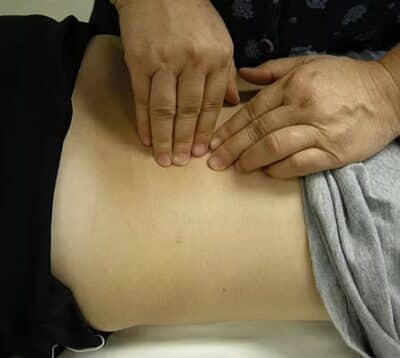
Connective Tissue Manipulation

Connective Tissue Manipulation is a technique using the soft pads of the fingers to move one layer of skin on the layer below. This reduces tension through the area being treated, typically the abdominal wall, the thighs and low back and gluteal areas. The reduction in tension around the blood vessel walls allows more blood to flow into the affected area and therefore can reduce inflammation. Softening the connective tissue also allows more movement to occur without causing irritation and therefore pain. The benefits of Connective Tissue Manipulation are cumulative. Once the tension has been lowered by treatment the reduction is maintained. Further reduction in connective tissue tension occurs with each additional treatment.
Visceral Manipulation
Visceral mobilization was used to treat women with generalized vulvadynia or provoked vulvadynia and it was found that there was:
*71% improvement in overall symptoms
*62% improvement in sexual function.
Visceral Manipulation (VM) was developed by world-renowned French Osteopath (DO) and Physical Therapist Jean-Pierre Barral. It evaluates and treats mobility of the internal organs, membranes fascia and ligaments of the body. Surgical scars, adhesion’s, illness, poor posture or injury causes straining on the connective tissues of the body. Visceral Manipulation can identify and treat these problems throughout the body. The technique is hand placement near the targeted structures creating tension to facilitate normal mobility of those areas. An example would be a person who presented with bladder pain, urgency and frequency may have increased tone of the bladder, urethra or pelvic floor muscles. If there is spasm of the bladder or urethra there is likely to be reflexive tightening of the surrounding pelvic floor muscles. Visceral mobilization treats the bladder to calm its tone down so the pelvic floor muscles can then also relax.
Core Stabilization

If you have ever taken a Pilates class or worked with an exercise trainer to stabilize your core, then you get the idea of spinal core stabilization. The goal is to strengthen the pelvic floor, spinal, gluteal and deep abdominal muscles to restore balance and strength at the pelvic base, which supports the rest of the trunk. A weak core can lead to bad posture, in turn putting stress upon joints, muscles and nerves, leading to inflammation and pain. Expect exercises to improve your core strength. Literature has shown that your small stabilizing muscles (transverse, abdominus, multifidus, pelvic floor and deep gluteal muscles) are the main stability of the lumbar spine and pelvis. It is important, to understand your body to be able to activate your deep core before engaging in Pilates, Barre, or yoga to get the most out of your practice and ensure proper stability
Joint Mobilization

Joint mobilization is a passive manual therapy technique applied to joints and their adjacent soft tissues that is used to normalize range of motion and decrease pain.
With this approach, the therapist applies pressure to the joints and surrounding soft tissue to return normal movement and reduce pain.
Joint mobilization can be a component of a treatment regimen for pelvic pain, low back pain, neck pain and headaches.
Myofascial Release
Myofascial release is a gentle, hands-on manual therapy technique that improves mobility, restores soft tissue and connective tissue flexibility, and increases ease of movement throughout the body.

Fascia is the connective tissue that surrounds and interconnects our muscles, bones, nerves, organs and vessels in a web of connective tissue. Connective tissues play several essential roles in our body and extend into every nook and cranny of the body interconnecting the body. All communication between cells occurs via the fascial web. Embedded in the fascia are millions of tiny nerves, blood vessels and mechanoreceptors that provide sensory and proprioceptive information to the brain.
The fascia is composed of collagen, elastin and polysaccharide gel, which form an interdependent matrix of strength, support, elasticity and shock absorption for the body. This allows the body to resist internal and external forms of mechanical stress.
Stress from injury, trauma, inflammation and even poor posture can result in thickening of collagen fibers resulting in fascial restrictions. These fascial restrictions can exert enormous pressure on the underlying structures resulting in pain, loss of motion and dysfunction of the system.
Myofascial release techniques can help alleviate these symptoms. The fascia responds to sustained pressure by decreasing the viscosity allowing for increased glide between structures. This results in increased blood flow and flexibility to the underlying tissue increasing nutrients to help heal the tissue.
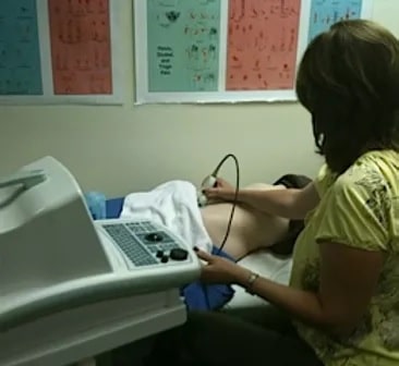
Rehabilitative Ultrasound Imaging (RUSI)

Rehabilitative Ultrasound Imaging (RUSI) has been used by research driven clinicians as a safe and cost effective method to enhance both the assessment and treatment of patients with motor control deficits of their lumbo-pelvic ‘core’ muscles, (transverse abdominis, lumbar multifidus, the diaphragm and the pelvic floor muscles). The value of Rehabilitative Ultrasound Imaging (RUSI) in a clinical setting is that it allows for real time study of these deep muscles as they contract. This allows both the patient and the therapist to view the contraction as it happens. Consequently, RUSI can be used as both an assessment tool, and more importantly as a form of biofeedback, providing patients with knowledge of performance in the early stages of motor relearning.
Soft Tissue Mobilization
Mechanical strains or injuries to the body can alter structure and function. Fascia (a layer of connective tissue that surrounds muscles, groups of muscles, nerves and blood vessels holding those structures together), muscles, and ligaments may tighten after an injury. Soft tissue mobilization techniques help evaluate and treat those areas that may not be moving correctly. There are 4 primary soft tissues of the body; skin, muscle, nerve and connective tissue. The term “mobilization” deals with the manipulation or movement of these tissues over joints that have restricted range of motion. These soft tissues around the bone need to be stretched in order to improve or restore range of motion and balance of the musculoskeletal system. Tightness in joints may restrict the flow of blood, lymph, and nerve signals in the area.

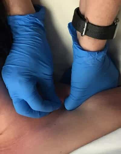
Functional Trigger Point Dry Needling or Intramuscular Manual Therapy (IMT)

Dry needling involves multiple advances of a filament needle into the muscle in the region of a “trigger point”. The aim of dry needling is to achieve a local twitch response to release muscle tension and pain. Dry needling is an effective treatment for chronic pain of neuropathic origin with very few side effects. This technique is unequaled in finding and eliminating neuromuscular dysfunction that lead to pain and functional deficits. The needle used is very thin and most subjects do not even feel it penetrate the skin. A healthy muscle feels very little discomfort with insertion of this needle. However, if the muscle is sensitive and shortened or has active trigger points within it, the subject will feel a sensation like a muscle cramp – “the twitch response”.
The patient also may feel a reproduction of their pain which is a helpful diagnostic indicator for the practitioner attempting to diagnose the cause of the patient’s symptoms. Patients soon learn to recognize and even welcome this sensation as it results in deactivating the trigger point, reducing pain and restoring normal length function to the involved muscle.
Surface EMG/Biofeedback
Surface electromyography (SEMG) biofeedback is a tool used to allow the patient to see how their pelvic floor muscles are working on a computer screen. The pelvic floor musculature is sometimes difficult to feel or to know if it is contracting and if it is contracting correctly. We use a surface electrode to place near the anal opening or an internal sensor placed either vaginally or rectally so the electrical activity of the pelvic floor can be monitored similar to an EKG used to look at cardiac function. The activity is then displayed on a computer screen to allow the patient to see and feel how the muscle functions. The use of EMG biofeedback allows one to isolate only the pelvic muscles and teaches the patient how to avoid using unwanted muscles like the buttock or abdominal muscles.
In 1996, US Department of Health and Human Services, Agency for Health Care Policy and Research (AHCPR), released an updated Clinical Practice Guideline or urinary incontinence, recommending that “behavioral procedures, such as biofeedback, be attempted before consideration of surgical or other invasive techniques.”
In a review of several studies using biofeedback to teach pelvic muscle exercises (Kegal’s exercises) for the treatment of incontinence (14), Tries states:
“…Patients benefit from biofeedback by developing a greater sense of control and mastery of bladder and bowel control, thus significantly reducing their fear, anxiety, isolation and hopelessness.”
1998 article in the Journal of the American Medical Association (JAMA) by Burgio reports:
“Patients treated with biofeedback showed a significantly greater reduction in urinary incontinence than a second group who received pharmaceutical intervention.”


